Carbon can exceed four-bond limit

A molecule originally proposed more than 40 years ago breaks the rules about how carbon connects to other atoms, scientists have confirmed. In this unusual instance, a carbon atom bonds to six other carbon atoms. That structure, mapped for the first time using X-rays, is an exception to carbon’s textbook four-friend limit, researchers report in the Jan. 2 Angewandte Chemie.

Although the idea for the structure isn’t new, “I think it has a larger impact when someone can see a picture of the molecule,” says Dean Tantillo, a chemist at the University of California, Davis who wasn’t part of the study. “It’s super important that people realize that although we’re taught carbon can only have four friends, carbon can be associated with more than four atoms.”
Atoms bond by sharing electrons. In a typical bond two electrons are shared, one from each of the atoms involved. Carbon has four such sharable electrons of its own, so it tends to form four bonds to other atoms.

But that rule doesn’t always hold. In the 1970s, scientists made an unusual discovery about a molecule called hexamethylbenzene. This molecule has a flat hexagonal ring made of six carbon atoms. An extra carbon atom sticks off each vertex of the ring, like six tiny arms. Hydrogen atoms attach to the ring’s arms. And leftover electrons zip around the middle of the ring, strengthening the bonds and making the molecule more stable.
When the scientists removed two electrons from the molecule, leaving it with a positive charge, some evidence suggested it might dramatically change its shape. It seemed to rearrange so that one carbon atom was bonded to six other carbons. But the researchers didn’t experimentally confirm that structure.
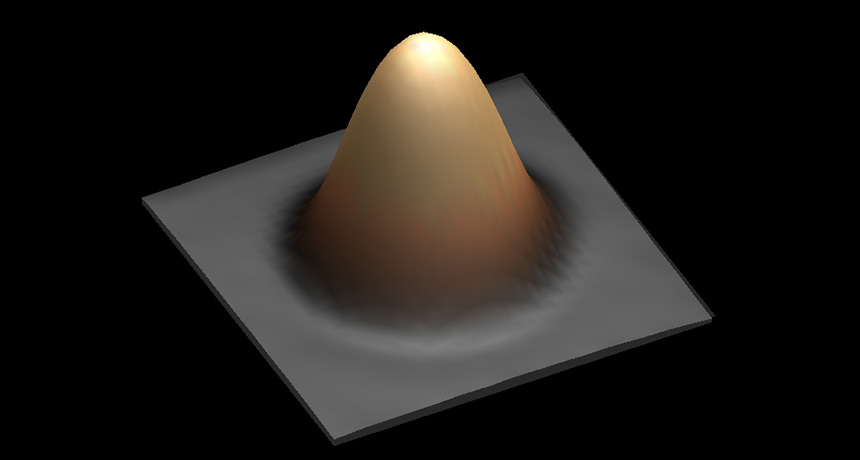
Now, a different lab has revisited the question. Making this charged version of hexamethylbenzene is a challenge because it’s stable only in extremely strong acid, says study coauthor Moritz Malischewski, a chemist at the Free University of Berlin. And the experimental details in the old study were a bit fuzzy. But after a bit of tinkering, he managed to create the charged molecule. He and coauthor Konrad Seppelt crystallized it with some other molecules, and then used X-rays to get a three-dimensional map of the crystal structure.

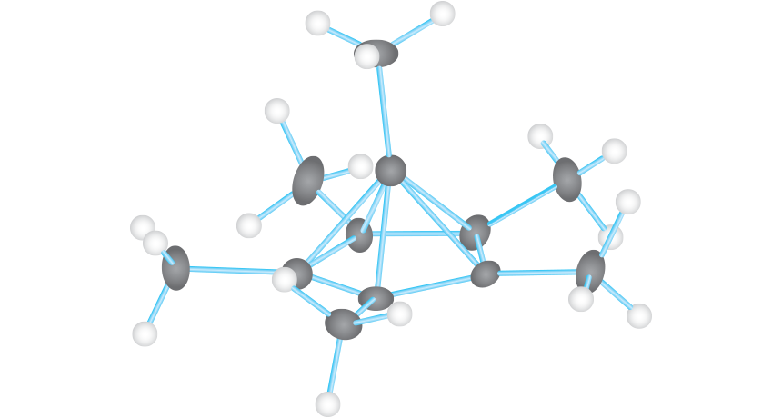
The X-ray experiment confirmed what other scientists had suggested in the 1970s: When hexamethylbenzene lost two electrons, it reordered itself. One carbon atom jumped out of the ring and took a new position on top, turning the flat hexagonal ring into a five-sided carbon pyramid. And the carbon on top of the pyramid was indeed bonded to six other carbons — five in the ring below, and one above.

“This molecule is very exceptional,” says Malischewski. Though scientists have found other exceptions to carbon’s four-bond limit, this is the first time carbon has been shown associating with this many other carbon atoms.

When Malischewski measured the length of the molecule’s chemical bonds, the top carbon’s six bonds were each a bit longer than an ordinary carbon-carbon bond. A longer bond is generally less strong. So by picking more partners, that carbon has a slightly weaker connection to each one.

“The carbon isn’t making six bonds in the sense that we usually think of a carbon-carbon bond as a two-electron bond,” Tantillo says. That’s because the carbon atom still has only four electrons to share. As a result, it spreads itself a bit thin by sharing electrons among the six bonds.

Bony head ornaments signal some supersized dinosaurs

Dinosaur fashion, like that of humans, is subject to interpretation. Bony cranial crests, horns or bumps may have served to woo mates or help members of the same species identify one another. While the exact purpose of this skull decor is debated, the standout structures tended to come with an even more conspicuous trait: bigger bodies.

Terry Gates, a paleontologist at North Carolina State University in Raleigh, and colleagues noticed an interesting trend in the fossil record of theropods, a group of dinosaurs that includes Tyrannosaurus rex and the ancestors of birds. Bigger beasts often sported skeletal headgear.
Across the family tree, Gates and his team analyzed 111 fossils dating from 65 million to 210 million years ago, and the trend held true. It makes sense: “Dinosaur size matters in terms of how they will be visually talking to one another,” says Gates. “When you’re smaller, your means of visual communication would be different than when you’re giant.”

The researchers also calculated that over time, theropod lineages with head ornaments evolved giant bodies (larger than 1,000 kilograms) 20 times faster on average than those without. Ornaments might have supersized some dinos, but researchers aren’t sure. The analysis, which appeared September 27 in Nature Communications, suggests theropods had to reach at least 55 kilograms to grow the headgear.

But among big-boned relatives of modern birds, skull toppers weren’t in vogue. Many of these dinos grew heavier than 55 kilograms, but they instead sported feathers that resembled those used by modern birds for flight. That might be because bigger, bolder feathers and showy headwear served similar ends. Gates speculates: “Once you have a signaling device in the form of a feather, why grow a bony cranial crest?” For these plumed dinosaurs, feathers were in and bony ornaments were out.

Size matters
Many large theropods, a group of dinosaurs that includes Tyrannosaurus rex and the ancestors of birds, had bony head ornaments such as crests, horns and bumps. New research suggests theropods had to reach at least 55.2 kilograms to grow the cranial decor. But big-boned dinos related to modern birds lacked the ornaments. Instead, they were decked out in feathers resembling those used by modern birds for flight.

Red Planet’s interior may not churn much

An enduring source of magma on Mars fueled volcanic eruptions for billions of years, clues inside a rock flung from the Red Planet reveal.

The newfound rock belongs to a batch of meteorites called shergottites that originated from the same Martian volcanic system, researchers report February 1 in Science Advances. But the new rock is considerably older than its counterparts. While previously discovered shergottites solidified from Martian magma between 427 million and 574 million years ago, the new rock formed around 2.4 billion years ago, chemical analyses show.
Such a wide range of ages means that a volcanic system on Mars churned out hot rocks from a stable source of magma for nearly half of the planet’s history, says study coauthor Thomas Lapen, a geologist at the University of Houston. That endurance could help scientists better understand Mars’ interior. “These are some of the longest-lived volcanoes in the solar system,” Lapen says.

Lapen and colleagues studied elements inside a Martian meteorite discovered in Algerian desert in 2012. Some of those elements serve as stopwatches that record the history of the rock. Isotopes of beryllium and aluminum, formed during exposure to cosmic rays, reveal that the rock zipped through space for around 1 million years. The steady decay of carbon 14 — left behind after cosmic ray collisions — suggests that the rock landed on Earth roughly 2,300 years ago. By combining these two measurements, the researchers found that the meteorite probably blasted off Mars alongside other shergottites a little over a million years ago. This exodus probably followed a massive impact in Mars’ volcano-filled Tharsis region.
The rocks share more than their exit route, the researchers found. Chemical similarities between the meteorites suggest that they all originate from the same source of hot rock deep within the Red Planet. That’s surprising given that the mix of radioactive elements inside the newfound meteorite suggests it solidified 1.8 billion years earlier than the next oldest shergottite, Lapen says.

Mars is known to have many volcanic systems across its surface, all fed by magma upwelling from the planet’s depths. Studies have previously suggested that some of these systems operated for billions of years. Though little is known about the Martian interior, many scientists had assumed that the magma feeding this volcanism changed over time as the Martian interior mixed. The absence of any difference in composition of the shergottites suggests Mars’ interior is relatively stagnant. That may result from Mars’ lack of plate tectonics, a process that helps blend Earth’s innards, Lapen proposes. Understanding the differences between Earth and Mars could help reveal why the two planets took such different trajectories, with Earth so much more life-friendly than Mars (SN: 5/2/15, p. 24).
Similarities between the shergottites could have another explanation, says planetary scientist Stephanie Werner of the University of Oslo. Large impacts can melt rocks, resetting their age. The shergottites may have formed around the same time billions of years ago before some had their ages altered by impacts over time, she proposes.

Upcoming missions will help illuminate what’s going on beneath the Martian surface, says James Head, a planetary scientist at Brown University in Providence, R.I. NASA’s InSight lander, currently slated for launch in 2018, will use seismic activity to map the Red Planet’s interior.

Observers caught these stars going supernova

In A.D. 185, Chinese records note the appearance of a “guest star” that then faded away over the span of several months. In 1572, astronomer Tycho Brahe and many others watched as a previously unknown star in the constellation Cassiopeia blasted out gobs of light and then eventually disappeared. And 30 years ago, the world witnessed a similar blaze of light from a small galaxy that orbits the Milky Way. In each case, humankind stood witness to a supernova — an exploding star — within or relatively close to our galaxy (representative border in gray, below).

Here’s a map of six supernovas directly seen by human eyes throughout history, and one nearby explosion that went unnoticed. Some were type 1a supernovas, the detonation of a stellar core left behind after a star releases its gas into space. Others were triggered when a star at least eight times as massive as the sun blows itself apart.

Common fungus may raise asthma risk

BOSTON — A fungus among us may tip the body toward developing asthma.

There’s mounting evidence that early exposure to microbes can protect against allergies and asthma (SN Online: 7/20/16). But “lo and behold, some fungi seem to put kids at risk for asthma,” microbiologist Brett Finlay said February 17 at a news conference during the annual meeting of the American Association for the Advancement of Science.

Infants whose guts harbored a particular kind of fungus — a yeast called Pichia — were more likely to develop asthma than babies whose guts didn’t have the fungus, Finlay reported. Studies in mice and people suggest that exposure to some fungi can both trigger and exacerbate asthma, but this is the first work linking asthma to a fungus in the gut microbiome of infants.
Finlay, of the University of British Columbia in Vancouver, and his colleagues had recently identified four gut bacteria in Canadian infants that seem to provide asthma protection. To see if infants elsewhere were similarly protected by such gut microbes, he decided to look at another population of children with an asthma rate similar to Canada’s (about 10 percent). He and his colleagues sampled the gut microbes of 100 infants in rural Ecuador and followed up five years later.

The researchers identified several factors that might influence risk of developing asthma, such as exposure to antibiotics, having respiratory infections, and whether or not the infants were breastfed. Of the 29 infants in the high-risk asthma group, more than 50 percent had asthma by age 5, Finlay said.

Surprisingly, the strongest predictor of whether a child developed asthma wasn’t bacterial. It was the presence of Pichia. And the yeast wasn’t protective; it tipped the scales toward asthma.

Finlay speculated that molecules made by the fungi interact with the infants’ developing immune systems in a way that somehow increases asthma risk. It isn’t clear how the infants’ guts acquire the fungus; some species of Pichia are found in soil, others in raw milk and cheese. Finlay and his colleagues are now going to look for the fungus in Canadian children’s gut microbes..

The researchers also looked at other gut microbe‒related factors that upped the Ecuadorean children’s asthma risk. Children with access to clean water had higher asthma rates, Finlay said. While drinking clean water helps people avoid several ills such as cholera, the link to asthma highlights how some dirt can be protective, he said. “We’ve cleaned up our world too much.”
This research underscores that caution should be used when generalizing about our intestinal flora. “What’s emerging is that it is very personalized,” gastroenterologist Eran Elinav of the Weizmann Institute of Science in Rehovot, Israel, said at the news conference. For example, evidence implicates some fungi in the development of inflammatory bowel disease, Elinav said, but it depends on the individual.

Scientists move closer to building synthetic yeast from scratch

Synthetic yeast is on the rise.

Scientists have constructed five more yeast chromosomes from scratch. The new work, reported online March 9 in Science, brings researchers closer to completely lab-built yeast.

“We’re doing it primarily to learn a little more about how cells are wired,” says geneticist Jef Boeke of the New York University Langone Medical Center. But scientists might also be able to tinker with a synthetic yeast cell more efficiently than a natural one, allowing more precise engineering of everything from antiviral drugs to biofuels.
Boeke was part of a team that reported the first synthetic yeast chromosome in 2014 (SN: 5/3/14, p. 7). Now, several hundred scientists in five countries are working to make all 16 Saccharomyces cerevisiae yeast chromosomes and integrate them into living cells. With six chromosomes finished, Boeke hopes the remaining 10 will be built by the end of 2017.

Each synthetic chromosome is based on one of S. cerevisiae’s, but with tweaks for efficiency. Researchers cut out stretches of DNA that can jump around and cause mutations, as well as parts that code for the same information multiple times.

When the researchers put chunks of synthetic DNA into yeast cells, the cells swapped out parts of their original DNA for the matching engineered snippets.

Yeast is a eukaryote — it stores its DNA in a nucleus, like human cells do. Eventually, this research could produce synthetic chromosomes for more complicated organisms, Boeke says, but such feats are still far in the future.

Single-atom magnets store bits of data

NEW ORLEANS — ­The tiniest electronic gadgets have nothing on a new data-storage device. Each bit is encoded using the magnetic field of a single atom — making for extremely compact data storage, although researchers have stored only two bits of data so far.

“If you can make your bit smaller, you can store more information,” physicist Fabian Natterer of the École Polytechnique Fédérale de Lausanne in Switzerland said March 16 at a meeting of the American Physical Society. Natterer and colleagues also reported the result in the March 9 Nature.
Natterer and colleagues created the minuscule magnetic bits using atoms of holmium deposited on a surface of magnesium oxide. The direction of each atom’s magnetic field served as the 1 or 0 of a bit, depending on whether its north pole was pointing up or down.

Using a scanning tunneling microscope, the scientists could flip an atom’s magnetic orientation to switch a bit from 0 to 1. To read out the data, the researchers measured the current running through the atom, which depends on the magnetic field’s orientation. To ensure that the change in current observed after flipping a bit was due to a reorientation of the atom’s magnetic field, the team added bystander iron atoms to the mix and measured how the holmium atoms’ magnetic fields affected the iron atoms.

The work could lead to new hard drives that store data at much greater densities than currently possible. Today’s technologies require 10,000 atoms or more to store a single bit of information.

Natterer also hopes to use these mini magnets to construct materials with fine-tuned magnetic properties, building substances a single atom at a time. “You can play with them. It’s like Lego,” he says.

For glass frogs, moms matter after all

Glass frogs often start life with some tender care from a source scientists didn’t expect: frog moms.

Maternal care wouldn’t be news among mammals or birds, but amphibian parenting intrigues biologists because dads are about as likely as moms to evolve as the caregiver sex. And among New World glass frogs (Centrolenidae), what little parental care there is almost always is dad’s job — or so scientists thought, says Jesse Delia of Boston University.
Months of strenuous nights searching streamside leaves in five countries, however, have revealed a widespread world of brief, but important, female care in glass frogs. In examining 40 species, Delia and Laura Bravo-Valencia, now at Corantioquia, a government environmental agency in Santa Fe de Antioquia, Colombia, found that often mothers lingered over newly laid eggs for several hours. By pressing maternal bellies against the brood, moms hydrated the jelly-glop of eggs and improved offspring chances of survival, Delia, Bravo-Valencia and Karen Warkentin, also of BU, report online March 31 in the Journal of Evolutionary Biology.

Glass frogs take their name from see-through skin on their bellies and, in certain cases, transparent organ tissues. (Some have clear hearts that reveal blood swishing through.) These frogs aren’t exactly obscure species, but until this field project, which stretched over six rainy seasons, female care in the family was unknown.

Female glass frogs may not cuddle their eggs for long, but it’s enough to matter, the researchers found. As is common in frogs, the mothers don’t drink with their mouths but absorb water directly through belly skin into a bladder. Moms pressing against a mass of newly laid eggs caused the protective goo to swell — perhaps by osmosis or peeing — and the mass to quadruple in size. For some of the glass frogs in the study, the youngsters were on their own once mom left. But at least hydration created an unpleasant amount of slime for a predator to bite through before getting to frog embryos.
Night-hunting katydids in captivity, when offered a choice, barely nibbled at a hydrated mass of frog offspring, concentrating instead on eating an unhydrated clutch. In the field, when researchers removed about two dozen moms from their clutches in two species, mortality at least doubled to around 80 percent. Predators and dehydration caused the most deaths.

There are still more than 100 glass frog species that Delia and Bravo-Valencia haven’t yet watched in the wild. But the researchers did track down maternal care in 10 of 12 genera. Such a widespread form of maternal care probably evolved in the ancestor of all glass frogs, the researchers propose after analyzing glass frog family trees several ways.

In contrast, prolonged care from glass frog dads — rehydrating the egg mass as needed and fighting off predators such as hungry spiders — seems to have arisen independently later, at least twice. Across evolution in the animal kingdom, “usually we don’t see transitions from female to male care,” Delia says. “The pattern we found is completely bizarre.”

Why females started hanging around their eggs at all fascinates Hope Klug, an evolutionary biologist at the University of Tennessee at Chattanooga who studies parental care. In frogs, with eggs mostly fertilized externally, females could easily leave any care to dad.

“Parental care is perhaps more common and diverse in animals than we realize,” she says. “We just might have to look a little bit harder for it.”

Toddlers’ screen time linked to speech delays and lost sleep, but questions remain

One of the most pressing and perplexing questions parents have to answer is what to do about screen time for little ones. Even scientists and doctors are stumped. That’s because no one knows how digital media such as smartphones, iPads and other screens affect children.

The American Academy of Pediatrics recently put out guidelines, but that advice was based on a frustratingly slim body of scientific evidence, as I’ve covered. Scientists are just scratching the surface of how screen time might influence growing bodies and minds. Two recent studies point out how hard these answers are to get. But the studies also hint that the answers might be important.

In the first study, Julia Ma at the University of Toronto and colleagues found that, in children younger than 2, the more time spent with a handheld screen, such as a smartphone or tablet, the more likely the child was to show signs of a speech delay. Ma presented the work May 6 at the 2017 Pediatric Academic Societies Meeting in San Francisco.

The team used information gleaned from nearly 900 children’s 18-month checkups. Parents answered a questionnaire about their child’s mobile media use and then filled out a checklist designed to identify heightened risk of speech problems. This checklist is a screening tool that picks up potential signs of trouble; it doesn’t offer a diagnosis of a language delay, points out study coauthor Catherine Birken, a pediatrician at The Hospital for Sick Children in Toronto.

Going into the study, the researchers didn’t have expectations about how many of these toddlers were using handheld screens. “We had very little clues, because there is almost no literature on the topic,” Birken says. “There’s just really not a lot there.”

It turns out that about 1 in 5 of the toddlers used handheld screens, and those kids had an average daily usage of about a half hour. Handheld screen time was associated with potential delays in expressive language, the team found. For every half hour of mobile media use, a child’s risk of language delay increased by about 50 percent.

“The relationship is not that strong,” Birken says, and those numbers come with big variations. Still, a link exists. And finding that association means there’s a lot more work to do, Birken says. In this study, researchers looked only at time spent with handheld screens. Future studies could investigate whether parents watching along with a child, the type of content or even time of day might change the calculation.

A different study, published April 13 in Scientific Reports, looked at handheld digital device use among young children and its relationship to sleep. As a group, kids from ages 6 months to 3 years who spent more time using mobile touch screen devices got less sleep at night.
Parent surveys filled out online indicated that each hour of touch screen use was linked to 26.4 fewer minutes of night sleep and 10.8 minutes more sleep during the day. Extra napping time “may go some way to offset the disturbed nighttime sleep, but the total sleep time of high users is still less than low users,” says study coauthor Tim Smith, a cognitive psychologist at Birkbeck, University of London. Each additional hour of touch screen use is linked to about 15 minutes less sleep over 24 hours.

By analyzing 20 independent studies, an earlier study found a similar link between portable screen use and less sleep among older children. The new results offer “a consistent message that the findings from older children translate into those younger,” says Ben Carter of King’s College London, who was a coauthor on the study of older children.

So the numbers are in. Daily doses of Daniel Tiger’s Neighborhood on a mobile device equals 7.5 minutes less sleep and a 50 percent greater risk of expressive language delay for your toddler, right? Well, no. It’s tempting to grab onto these numbers, but the science is too preliminary. In both cases, the results show that the two things go together, not that one caused the other.

It may be a long time before scientists have answers about how digital technology affects children. In the meantime, you can follow the American Academy of Pediatrics’ recently updated guidelines, which discourage screens (except for video chatting) before 18 months of age and for all children during meals or in bedrooms.

We now live in a world where smartphones are ever-present companions, a saturation that normalizes the sight of small screens in tiny hands. But I think we should give that new norm some extra scrutiny. The role of mobile devices in our kids’ lives — and our own — is something worth thinking about, hard.

Petunias spread their scent using pushy proteins

When it comes to smelling pretty, petunias are pretty pushy.

Instead of just letting scent compounds waft into the air, the plants use a particular molecule called a transporter protein to help move the compounds along, a new study found. The results, published June 30 in Science, could help researchers genetically engineer many kinds of plants both to attract pollinators and to repel pests and plant eaters.

“These researchers have been pursuing this transporter protein for a while,” says David Clark, an expert in horticultural biotechnology and genetics at the University of Florida in Gainesville. “Now they’ve got it. And the implications could be big.”
Plants use scents to communicate (SN: 7/27/02, p. 56). The scent compounds can attract insects and other organisms that spread pollen and help plants reproduce, or can repel pests and plant-eating animals. The proteins found in the new study could be used to dial the amount of scent up or down so that plants can attract more pollinators or better protect themselves. Currently unscented plants could be engineered to smell, too, giving them a better shot at reproduction and survival, Clark says.
Plants get their scents from volatile organic compounds, which easily turn into gases at ambient temperatures. Petunias get their sweet smell from a mix of benzaldehyde, the same compound that gives cherries and almonds their fruity, nutty scent, and phenylpropanoids, often used in perfumes.

But nice smells have a trade-off: If these volatile compounds build up inside a plant, they can damage the plant’s cells.
About two years ago, study coauthor Joshua Widhalm, a horticulturist at Purdue University in West Lafayette, Ind., and colleagues used computer simulations to look at the way petunias’ scent compounds moved. The results showed that the compounds can’t move out of cells fast enough on their own to avoid damaging the plant. So the researchers hypothesized that something must be shuttling the compounds out.

In the new study, led by Purdue biochemist Natalia Dudareva, the team looked for genetic changes as the plant developed from its budding stage, which had the lowest levels of volatile organic compounds, to its flower-opening stage, with the highest levels. As flowers opened and scent levels peaked, the gene PhABCG1 went into overdrive; levels of the protein that it makes jumped to more than 100 times higher than during the budding stage, the researchers report.

The team then genetically engineered petunias to produce 70 to 80 percent less of the PhABCG1 protein. Compared with regular petunias, the engineered ones released around half as much of the scent compounds, with levels inside the plant’s cells building to double or more the normal levels. Images of the cells show that the accumulation led to deterioration of cell membranes.

A lot of work has been done to identify the genes and proteins that generate scent compounds, says Clark. But this appears to be the first study to have identified a transporter protein to shuttle those compounds out of the cell. “That’s a big deal,” he says.